Marmoset MRI Technical Notes
Magnetic resonance imaging (MRI) is a method to visualize the information obtained by nuclear magnetic resonance of Hydrogen (mainly water in vivo) using magnetism and microwave. MRI can acquire images in any directions, free from irradiation of X-ray , although it has relatively long imaging time and produces loud scanning noise. MRI machines are broadly used in clinics mainly for medical imaging diagnosis, and more than five thousand MRI scanners have been actively installed in hospitals and research institutes in Japan. MRI can provide high contrast and spatial resolutions, applied to image not only the brain but also the body and limbs. MRI use a very
strong magnetic field, and 3T (=30000 gauss) is common in clinics.
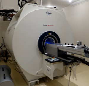
The current project uses a non-human primates, common marmoset, which is much smaller than humans. Therefore, we use 9.4T MRI that achieves the spatial resolution of hundred micrometers for obtaining an image slice in vivo. This imaging technique is highly valuable in experimental medicine as it makes a significant contribution to reduction and refinement in the three Rs of animal ethics. Moreover, the same imaging protocols can be applicable regardless of the strength of magnetic field, and therefore it would be straightforward to transfer research techniques and methods developed in 9.4T MRI to those for 3.0T MRI in clinical applications.
[DataID: 2406, Creator: Okano Lab]