Marmoset Tracer Projects
Marmoset brain mapping based on tracer injection
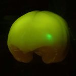
Brain/MINDS is making a connection map of the marmoset cortex using anterograde tracer injection with virus vectors. In the first phase of the project (2014-2018), three groups, led by Tetsuo Yamamori, Noritaka Ichinohe, and Partha Mitra, were involved. The Yamamori team used serial two-photon tomography (STPT) for imaging the projections while Ichinohe and Mitra groups both used a digital slide scanner. For accurate 3D registration, all of the tracer-injected brains were imaged with an MRI-scanner before sectioning. The tracer signals were mapped to a reference 3D space, which will be used for integration of cross-modal experiments, including MRI, DTI and histological staining, in addition to the tracer data. In the second phase of the project (2019-2023) the tracer injection work has been consolidated and is continued on by Yamamori Team.
Yamamori Team, RIKEN CBS
Ichinohe Team, National Institute of Neuroscience
Mitra Team, Cold Spring Harbor Laboratory
Features
-
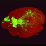
PFC Projection: 3D Movie
An example movie to demonstrate the axonal projections from the prefro...
-

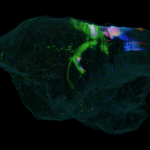
TL Projection: 3D Movie
A sample of the 3D reconstructed marmoset atlas from tracer images.
-
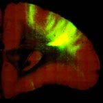
PL Projection: 3D Movie
The marmoset brain with tracer injection to A46 was processed by seria...
-
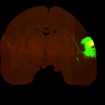
PFC Projection: Section Images
Image stacks for coronal sections of PFC projections
-
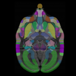
TL Projection: Section Images
Axonal projection maps in the whole marmoset brain.
-
PL Projection: Section Images
Fully interactive portal site with user friendly options. Ability to n...